
V8 General Imaging
The V8 ultrasound system combines exquisite imaging quality powered by Crystal Architecture™ with efficient, streamlined examination enabled by Intelligent Assist tools, and reengineered workflow to fulfill the needs of today's busy clinical environment.
.png)
Redefined imaging technologies powered by Crystal Architecture™
Crystal Architecture™, an imaging architecture that combines CrystalBeam™ and CrystalPure™,while based upon S-Vue Transducer™, is to provide crystal clear image.
-
CrystalBeam™ is a new beamforming technology beneficial in delivering high-quality image resolution and increased uniformity of images.
-
CrystalPure™ is Samsung’s up-to-date ultrasound imaging engine with enhanced 2D image processing, color signal processing, and advanced intellectual algorithm to offer outstanding image performance and efficient workflow during complex cases.
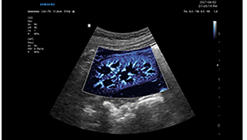
Exquisite imaging qualityfor reliability and confidence
Gain insight into the problem based on exceptional image performance powered bySamsung’s core imaging engine, Crystal Architecture™. The premium imaging engine combinesthe benefits of enhanced 2D image processing and detailed expression of color signal processing.
.png)
Intercostal view / Intercostal view with ShadowHDR™
ShadowHDR™ selectively applies high-frequency and low-frequency of the ultrasound to identify shadow areas such as fetal head or spine where attenuation occurs.
.png)
Quadriceps tendon / Quadriceps tendon with HQ-Vision™
HQ-Vison™ provides clearer images by mitigating the characteristics of ultrasound images that are slightly blurred than the actual vision.
.png)
Biceps tendon / Biceps tendon with ClearVision
ClearVision The noise reduction filter enhances the edge contrast and creates sharp 2D images for optimal diagnostic performance. In addition, ClearVision provides application-specific optimization and advanced temporal resolution in live scan mode.
.png)
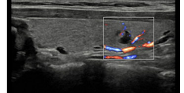
Thyroid Nodule with S-Flow™
S-Flow™, a directional Power Doppler imaging technology, can help to detect even the peripheral blood vessels. It enables accurate diagnosis when the blood flow examination is especially difficult.
.png)
Kidney with MV-Flow™
MV-Flow™ ¹ offers an advanced color imaging for visualizing slow flow of microvascularized structures. High frame rates and advanced filtering enable MV-Flow™ to provide a detailed view of blood flow in relation to surrounding tissue or pathology with enhanced spatial resolution.
.png)
Thyroid nodule (S-Flow™ with LumiFlow™)
LumiFlow™ ¹ is a function that visualizes blood flow in three dimensional-like to help understand the structure of blood flow and small vessels intuitively.
Intelligent Assist toolsfor efficient examination
Simplify operation and enhance diagnostic confidence with built-in Intelligent Assist features.V8 supports healthcare professionals with semi-automated features they need to help making decisions. The system is equipped with a range of tools that help accurately diagnose issues and achieve greater throughput.
.png)
Display and quantify tissue stiffness in a non-invasive method
S-Shearwave Imaging™ ¹ allows for non-invasive assessment of stiff tissues in various applications. The color-coded elastogram, quantitative measurements, display options, and user-selectable ROI functions are especially useful for accurate diagnosis of breast and liver diseases.
.png)
Quantify wall motion of the left ventricle
Strain+ ¹ is a quantitative tool for measuring global and segmental wall motion of the left ventricle (LV). In Strain+, three standard LV views and a Bull's Eye are displayed in a quad screen for easy and quick assessment of the LV function.
.png)
Detect functional changes of cardiovascular vessels
ArterialAnalysis™ ¹ detects functional changes of vessels, providing measurement values such as the stiffness, intima-media thickness and pulse wave velocity of the common carotid artery. Since the functional changes occur before morphological changes, this technology supports the early detection of cardiovascular disease.
Perform multi-modality fusion biopsies with high precision
S-Fusion™ ¹ enables simultaneous localization of a lesion using real-time ultrasound with other volumetric imaging modalities, enabling accurate targeting during interventional and other advanced clinical procedures. Samsung's new Auto Registration helps quickly and precisely fuse the images for increased efficiency.
Contrast Enhanced Ultrasound
CEUS+ ¹ is a contrast enhancement imaging technology that utilizes the characteristics of ultrasound contrast agents. The microbubble contrast agent injected into the body through the vein or alike is subjected to perform nonlinear resonance due to stimulation of ultrasound energy. In addition to the nonlinear signal generated by this method, the ultrasound contrast image is implemented by using the harmonic signal and thus utilized for the diagnosis based on the contrast characteristics over time.
Score and report wall motion to determine heart and blood vessel function
StressEcho ¹ package includes wall motion scoring and reporting. It includes exercise StressEcho, pharmacologic StressEcho, diastolic StressEcho and free programmable StressEcho.
Measure IMT in one click
AutoIMT¹ is a screening tool to analyze a patient's potential risk of cardiovascular disease. It allows easy intima-media thickness measurement of both the anterior and posterior wall of the common carotid by the click of a button.
Re-engineered workflow and design for a simplified process
Ease your day by streamlining workflow with V8's convenient features and collaborative solutions that reduce multiple tasks into just a few steps and keystrokes. How we display the scan data more easily and precisely is an important focus for the user experience. The ergonomic design makes effective use of the user’s working environment to assure utility and productivity.
.png)
.png)
.png)